Sciatica
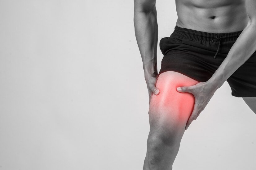
Sciatica is a term used for pain that radiates from the lower back down to the legs. The most common cause of sciatica is irritation or tightness of the muscles that run from the back down to the legs. It may be accompanied by weakness, trembling, and wasting in the distribution of the affected muscles.
Common minimally invasive non surgical interventions I offer in pain clinic for knee pain include

A nerve root block is used for people suffering from radicular pain (in simple terms sciatica or pain radiating from the spine to the legs). It helps deliver maximum drug close to the area of actual pathology- disc bulge/ nerve compression with a favorable response especially if the procedure is performed soon after the onset of symptoms. It involves injection close to the nerves as they come out of the spine. A needle is placed under x-ray guidance and a dye (contrast agent) is given to check needle position prior to giving the local anaesthetic and steroid mixture. These injections can provide diagnostic information and therapeutic benefits.
Epidural space is present in the spine around the sac containing the spinal cord and the nerves. It extends from the back of the head to the bottom of spine. Epidural injection involves placing a needle in this space under x-ray guidance. A dye (contrast agent) is used to confirm needle placement before a mixture of local anaesthetic and steroid is given. The level at which the injection is performed will depend on the actual pathology site and the pain distribution.
Depending on the level at which the epidural injection is performed, it may be termed as
- Cervical epidural – Indicated for neck and arm pain. It involves performing an injection at the base of neck under x-ray guidance.
- Thoracic epidural - Indications for this injection include mid back, chest or abdominal pain. It is performed at mid back level between the shoulder blades, under x-ray guidance.
- Lumbar epidural is performed for back, groin or leg pain. It involves a x-ray guided injection in the lower back at the waist level.
- Caudal epidural is performed for back and leg pain. It involves a x-ray or ultrasound-guided injection close to the lower end of the spine near the tailbone.
Spine has many vertebrae and these are linked to each other by small joints called facet joints. The main function of these joints is to provide stability while allowing some degree of movement. These joint commonly become painful and stiff as a result of wear and tear, inflammation or injury. The resulting pain is generally described as a dull ache, heaviness that may radiate towards buttock and thigh.
Investigations such as x-rays and MRI may or may not show joint changes. It is important to understand that even if these investigations show wear and tear/arthritis, not every arthritic joint is painful so MRI findings alone cannot be relied on to make the diagnosis. A more reliable test to determine if these joints are responsible for your back pain is accurately placed injections and if the pain reduced significantly then these joints are the likely source of pain.
Injections for facet joints can be done under x- ray or ultrasound guidance as a day care procedure. The procedure involves placing needles at precise location under x-ray guidance followed by injection of a mixture of local anaesthetic and steroid. Most people tolerate this procedure well under local anaesthesia. It may take a few days and sometimes weeks for the full effects of the injections to become apparent.
Medial branch Blocks
These injections are used as tests to diagnose facet joint pain and assess whether the radiofrequency treatment will be beneficial or not. The targets in these injections are the nerve carrying the pain sensation from the facet joints (compared with the joints themselves in the facet joint injections). The procedure involves placing small amount of local anaesthetic at specific locations under x-ray guidance. The resulting nerve block temporarily abolishes the pain if the source of pain is the facet joints. Decision to proceed with radiofrequency is taken based on the degree and duration and of pain relief obtained from these diagnostic injections.
Lumbar sympathetic nerves run on either side of the spine and are responsible for controlling blood supply to the legs. Sometimes these nerves get involved in carrying pain sensations and contribute to persisting pain.
Lumbar sympathetic block interrupts the flow of signals in these nerves and produces pain relief and increased blood flow to legs as a consequence. These injections can help in conditions with reduced blood supply to legs such as ischemic leg pain, non healing leg ulcers and other pain conditions involving the sympathetic nerves such as in Complex Regional Pain Syndrome, post amputation phantom limb pain.
Block can be performed for diagnostic or therapeutic reasons. The difference between the two is in the drug that is injected. Diagnostic blocks involve injection of local anaesthetics to establish whether or not these nerves are involved in carrying the pain sensation. The relief from diagnostic injections may be short lasting, nevertheless it has an important contribution towards establishing further course of action and provides an opportunity to interrupt the pain cycle and engage in physical therapy. Therapeutic injections are aimed at providing prolonged pain relief.
Piriformis and Obturator internus are buttock muscles in close proximity to one of the major leg nerves (sciatic nerve) as it leaves the pelvis and enters the leg. Pressure on this nerve can lead to sciatica/ nerve pain. This usually presents as buttock and leg pain which is worse in sitting position.
Piriformis muscle extends from the side of the sacrum, tailbone to the upper part of the thigh bone (femur). Spasm, swelling or irritation of muscle can cause buttock pain and irritation of the sciatic nerve. Indications for injections include diagnostic purposes or for providing pain relief (therapeutic). It is performed under ultrasound guidance and involves injecting a mixture of local anaesthetic and steroid.
Skeletal Muscles form a substantial proportion of human body and their ability to contract and relax helps in producing body movements. When muscles fail to relax, they form knots or tight bands known as trigger points. These can be a result of inflammation, trauma and injury of the muscle or the neighbouring structures. Poor posture and repetitive strain are other predisposing factors. They are more commonly observed in trapezius, neck and lower back muscles. Pressure over a trigger point produces local soreness and may refer pain to other body parts. Trigger points can limit the range of movement; affect posture predisposing other areas to unaccustomed strain.
Trigger point injections are performed in an outpatient/ day-care setting and involve injection of a mixture of local anaesthetic and steroid. I prefer to perform these injections under ultrasound guidance as this helps in improving the accuracy and reduces the chances of complications. Post injection physiotherapy is essential to prevent recurrence and maximise the benefits.
Hamstrings are a group of muscles present at the back of thigh. They extend from the pelvis (ischial tuberosity) to the knee and play an important role is everyday activities such as bending or running. Muscles attach to the bones with the help of a special type of tissue called tendons. With overuse, misuse or injury, these tendons can get inflamed, torn leading to development of a condition called tendinopathy. When the proximal part of hamstrings is involved it presents as buttock pain radiating down the back of knee. Pain may be worse on sitting on a firm seat. Sciatic nerve is present close by and its irritation can cause pain to radiate further down the leg.
Condition such as unequal leg length, core and pelvic muscle weakness, being overweight and repeated overloading with insufficient warm up predispose to development of hamstring tendinopathy. Higher incidence is seen in runners, football players, dancers and older adults who do a lot of walking.
Treatment options include rest, activity modification, physical therapy and medications. If these fail to produce desired results then injections with Platelet Rich Plasma (PRP), autologous blood (ABI) or steroids are considered. Percutaneous tenotomy is another option. Injections are performed under ultrasound guidance in the peritendinous region. Direct injection into the tendons is avoided. These are performed under local anaesthesia as an outpatient procedure or a day case.
Trochanteric Bursitis presents as pain on the outer side of the hip joint. It now been renamed as Greater Trochanteric Pain Syndrome (GTPS) as a range of conditions including muscle tears, tendon issues and trigger points can produce similar symptoms. This region houses greater trochanter (bony edge of the top of the thigh bone) and numerous burase (to prevent friction). A number of buttock muscles attach of the greater trochanter.
GTPS is more common in middle aged or elderly women and in athletes involved in prolonged running. Trauma, repetitive stress, leg length discrepancies and weak hip muscles can predispose to developing this condition. It presents as pain on the outer side of hip which can radiate towards the knee. Lying on the affected side may be uncomfortable.
Conservative treatment involves avoidance of activities that aggravates the problem, ice, weight management, painkillers and physiotherapy. Injections are considered if conservative management is not producing desired results. Local anaesthetic and steroid injection can help break the pain cycle and facilitate physical therapy. These are performed under ultrasound guidance to ensure delivery of medication at the correct site and reduce changes of complications.

